Suprabhat Healthcare is a fully registered Corneal Transplant Centre recognized by the state health authorities. Various corneal microsurgical procedures are carried out in complex cases.
Dr Prashant Bhartiya has been trained at the premier institute of India in Ophthalmology at the R P Centre for Opthalmic Sciences, All India Institute of Medical sciences. He has a fellowship in Cornea and refractive Surgery from the prestigious Royal Victorian Eye and Ear Hospital at Melbourne, Australia. Dr Prashant has to his credit performing the first successful DSEK of central India in 2010. He has various papers on Surgical techniques with two successive best video awards at the American Association of Ophthalmology annual conferences. His technique in managing difficult situations after DALK has received the Dr H C Sethiya award at the annual conference of the Madhya Pradesh State Ophthalmic society.
A corneal transplantation involves replacing a diseased, scarred, or cloudy cornea with a healthy one from a donor. Dr Prashant Bhartiya does specialize in several different techniques mentioned below.
• Penetrating Keratoplasty: This transplant procedure replaces the entire cornea with donor tissue. Sometimes called a full-thickness transplant, this procedure may be performed in patients with advanced keratoconus, Traumatic or post infectious scars, or pseudophakic bullous keratopathy.
• Deep Anterior Lamellar Keratoplasty (DALK): Unlike PK, this partial-thickness transplant procedure preserves the patient’s Descemet’s membrane and endothelium. This procedure may be useful for conditions that affect the thickest layer of the cornea, called the corneal stroma, as long as the endothelium is healthy.
• Endothelial Keratoplasty or Descemets Stripping Endothelial Keratoplasty (DSEK) and Descemets Membrane Endothelial Keratoplasty (DMEK): In Endothelial Keratoplasty, the innermost layer of the cornea, known as the endothelium, is replaced. It is often performed in patients who have advanced pseudophakic bullous keratopathy or Fuchs’ endothelial dystrophy.
In corneal transplantation, the aim is to replace a diseased, scarred, or cloudy cornea with a healthy one in order to allow light to reach the retina (similar to the film of a camera) in the back of your eye. Special surgical instruments are used to remove the diseased part of the cornea and replace it with a clear donor cornea. Fine sutures secure the donor cornea in place, and antibiotic drops are given to prevent infection. The eye is dressed with sterile gauze pads and a protective shield. Following surgery, the eye needs to be protected and eye drops should be applied for several months to years to promote healing and prevent rejection (failure of the transplant). Some patients get good vision in two to three months, while others must wait about a year for complete healing.
Corneal transplants have restored sight to many people who would have been blinded permanently by corneal injury, infection, or inherited corneal disease or degeneration. The most important factors determining success are the underlying disease process and the quality of the tissue used during transplantation. In some cases, the body may start to reject the new cornea. If this happens, medication can be administered at the first sign of symptoms, which increases the chance of success. For this reason, it is important that patients immediately report sudden changes in their condition to their surgeon. There are four signs of rejection that can be remembered by the mnemonic RSVP: Redness, Sensitivity to light, decreased Vision, or Pain.
Suprabhat Healthcare is one of a few hospitals in Indore that performs the procedure called corneal cross-linking. This minimally invasive, in-office procedure is the only treatment that can slow or stop the progression of keratoconus—an eye condition that causes the cornea to become thin, weak, and irregularly shaped. It can also be used to treat rare complications from LASIK surgery.
Corneal cross-linking offers new hope for patients with Keratoconus. Previously, the only treatment options available were eye glasses/contact lenses and, in advanced cases, corneal transplant surgery. Now with corneal cross-linking, we can preserve vision and prevent the condition from worsening.
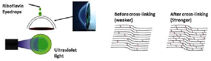
Corneal collagen cross-linking, sometimes called CXL or C3R, helps strengthen the cornea. During the procedure, liquid vitamin B2 (riboflavin) is placed in the eyes and exposed to ultraviolet light. This treatment increases the collagen fiber cross-linkages, which act like the cornea’s support beams—making the cornea stronger and increasing its ability to maintain its shape and focusing power.
There are risks (problems) that can happen with C3R. Here are some of the most common or serious:
• Pain. Usually relieved with pain killers required during first few days.
• Infection. You could get an eye infection from the surgery especially if you do not take adequate precautions to prevent dust and dirty water from entering the eye.
• Vision and Healing issues: a little corneal scarring is expected from the procedure. It may be severe in some cases and may decrease the vision. A corneal transplant or Contact lens may be required to improve vision later.
The goal of CXL is to stop the cornea from getting thinner, weaker, and more irregular in shape. Although CXL cannot make your cornea normal again, it may keep your vision from getting worse. Sometimes, your vision may even improve. But you may still need to wear glasses or contact lenses for near or far vision. It is possible that your eye may start getting weak again. If it does, you may need another CXL treatment or another type of cornea surgery.
The most common protocol used at Suprabhat Healthcare is the Dresden Protocol as it is reported as the most successful and safe.
Amniotic membrane is a thin tissue that lines the amniotic sac during a woman’s pregnancy. The function of the amniotic membrane is to provide a protective environment for the baby to develop. Many scientists believe that the amniotic membrane closely resembles the tissues of the outer lining of the eye and contains cells that can assist in healing and pain management.
Amniotic Membrane has three main aims in clinical use:
• To promote healing (Epithelialization)
• To reduce pain
• To minimize inflammation of the ocular surface.
This allows procedures to be performed in a sequential manner, such as cornea transplantation with a reduced risk of rejection.
Amniotic Membrane is available at Suprabhat healthcare and various types of surgeries are carried out which include use of Amniotic Membrane Transplantation.
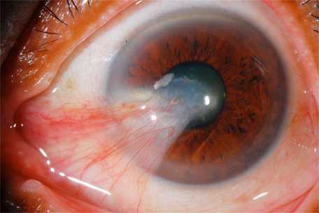
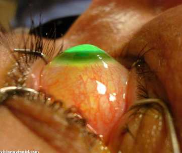
Pterygium surgery involves removal of the abnormal tissue from the sclera and cornea of the eye. Today’s techniques offer a significantly higher success rate than conventional surgery.
In traditional “bare sclera” pterygium removal, the underlying white of the eye is left exposed. Healing occurs over two to four weeks with mild to moderate discomfort. Unfortunately, “bare sclera” pterygium surgery has a high rate of re-growth; this occurs in up to 50% of patients. In many cases, the pterygium grows back larger that its original size.
Most corneal specialists today perform pterygium surgery with a conjunctival autograft because of a reduced risk of recurrence. In this technique, the pterygium is removed, and the cornea regains clarity. However, the gap in the mucous membrane (conjunctiva) tissue, where the pterygium was removed, is filled with a transplant of tissue that has been painlessly removed from underneath the upper eyelid. Although the procedure requires more surgical skill than traditional surgery, this “auto-graft” (self-transplant) helps prevent re-growth of the pterygium by filling the space where abnormal tissue would have re-grown.
• Stem Cell Transplantation, SLET
Special surgical technique has been evolved which helps in restoration of the ocular surface in cases with extensive ocular burns, chemical injuries or diseses like stevens Johnson syndrome.
• Glueing
Early application of cyanoacrylate glue with bandage contact lens is known to aid management of small corneal perforations, corneal melts and wound leaks. It may also help in avaiding the need for tectonic keratoplasty.
• Corneal Tattoing

Cosmetic tattoing of corneal opacities is possible at Suprabhat Healthcare. Various options for place the dark coloured dye is available and can be helpful in covering corneal whiteness.
© 2018 Suprabhat Healthcare. All rights reserved.